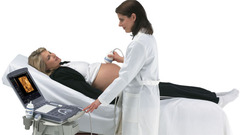
4D Real Time
4D is shorthand for “four-dimensional” -- the fourth dimension being time. As far as ultrasound is concerned, 4D Ultrasound is the latest ultrasound technology. 4D Ultrasound takes three-dimensional ultrasound images and adds the element of time to the process. The result -- live-action images of your unborn child.
VOCAL II VCI (Volume Contrast Imaging)
A very thin volume 4D scan with some minor differences in mechanics of acquisition is called as VCI (volume contrast imaging) VCI increases the inherent contrast in the image and delineates borders in a better manner.SonoVCAD labor

Sonography-based Volume Computer Aided Display labor (SonoVCAD™ labor) is a proprietary 3D automated tool that allows you to confidently measure fetal head progression, rotation and direction while automatically documenting the labor procedure with objective ultrasound and manual data in one easy report.
DICOM 4D
DICOM (Digital Imaging and Communication in Medicine) is a standard that describe the way medical information are stored/exchanged in the radiological department. The purpose of DICOM is to facilitate interaction between medical devices.Digital technology has in the last few decades entered almost every aspect of medicine. There has
been a huge development in noninvasive medical imaging equipment. Because there are many medical
equipment manufacturers, a standard for storage and exchange of medical images needed to be developed.
DICOM (Digital Imaging and Communication in Medicine) makes medical image exchange more easy and
independent of the imaging equipment manufacturer. Besides the image data, DICOM file format supports other information useful to describe the image.
STIC [Spatio-Temporal Image Correlation]
In STIC technology, an automated device incorporated into the ultrasound probe performs a slow sweep acquiring a single threedimensional (3D) volume. This volume is composed of a great number of two-dimensional (2D) frames. As the volume of the fetal heart is small, appropriately regulating the region of interest allows a very high frame rate (in the order of 150 frames per second) during 3D volume acquisition. For example, an acquisition time of 10 seconds and a sweep angle of 25° would lead to the
recording of 1500 2D frames. During the acquisition time there will be approximately 20-25 cardiac cycles. Therefore, among the 1500 frames, 20-25 will show a systolic peak
recording of 1500 2D frames. During the acquisition time there will be approximately 20-25 cardiac cycles. Therefore, among the 1500 frames, 20-25 will show a systolic peak


No comments:
Post a Comment